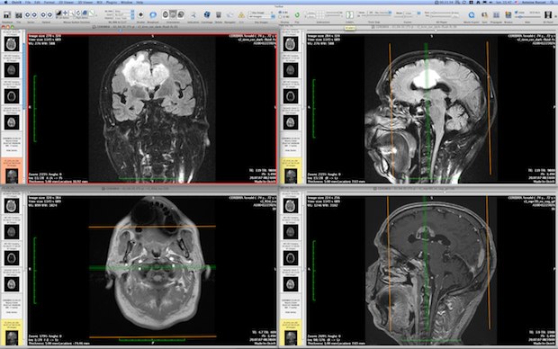

All these modes support 4D data and are able to produce image fusion between two different series (PET-CT and SPECT-CT display support). The 3D Viewer offers all modern rendering modes: Multiplanar reconstruction (MPR), Surface Rendering, Volume Rendering and Maximum Intensity Projection (MIP). OsiriX has been specifically designed for navigation and visualization of multimodality and multidimensional images: 2D Viewer, 3D Viewer, 4D Viewer (3D series with temporal dimension, for example: Cardiac-CT) and 5D Viewer (3D series with temporal and functional dimensions, for example: Cardiac-PET-CT). OsiriX is able to receive images transferred by DICOM communication protocol from any PACS or imaging modality (C-STORE SCP/SCU, and Query/Retrieve : C-MOVE SCU/SCP, C-FIND SCU/SCP, C-GET SCU/SCP, WADO). It is fully compliant with the DICOM standard for image comunication and image file formats.
#FILE FORMAT OF AN OSIRIX DICOM DATA SET SOFTWARE#
OsiriX is an image processing software dedicated to DICOM images (“.dcm” / “.DCM”extension) produced by imaging equipment (MRI, CT, PET, PET-CT, SPECT-CT, Ultrasounds, …). The tool is designed especially for neurobiologists, and it helps them better visualize the fluorescent-stained confocal samples. It combines the renderings ofmulti-channel volume data and polygon mesh data, where the properties of each dataset can be adjusted independently and quickly. Visualization Software FluoRenderįluoRender is an interactive rendering tool for confocal microscopy data visualization. We list some of them here for easy reference, along with brief descriptions.


There are a number of freely-available tools from other National Research Resources and elsewhere. CISMM is one of several groups providing visualization and analysis tools freely usable by the biomedical microscopy community.


 0 kommentar(er)
